| dc.contributor.advisor | Ahluwalia, Balpreet S. | |
| dc.contributor.advisor | Basnet, Purusotam | |
| dc.contributor.advisor | Wolfson, Deanna L. | |
| dc.contributor.author | Singh, Rajwinder | |
| dc.date.accessioned | 2018-01-02T13:08:13Z | |
| dc.date.available | 2018-01-02T13:08:13Z | |
| dc.date.issued | 2017-05-15 | |
| dc.description.abstract | Approximately 800 women die from pregnancy or childbirth-related pregnancy complications around the world every day. One of the major reasons for these complications is inflammation following infection. Approximately 40% cases of preterm births occur due to microbial invasion in the genitourinary track. During inflammation, macrophages present at the feto-maternal junction release an increased amount of nitric oxide (NO) and other important pro-inflammatory cytokines: TNF-alpha and INF-gamma. This can disturb the trophoblast function which can lead to pregnancy complications, such as abortion, pre-eclampsia and birth defects.
Here I aimed to study the cellular and sub-cellular morphological changes in macrophages (RAW264.7) and trophoblasts (HTR-8/SVneo) following externally supplied inflammatory agents: lipopolysaccharides (LPS), concanavalin-A (Con-A) and tumor necrosis factor alpha (TNF-alpha), in vitro.
Nitric oxide (NO) produced by the cells under inflammatory response was measured quantitatively using the Griess reagent test. Morphological changes were examined in response to LPS, Con-A and TNF-alpha challenge on live macrophages and trophoblasts using structured illumination microscopy (SIM) and quantitative phase microscopy (QPM).
LPS-challenged macrophages produced approximately 22 folds more NO as compared to controls, whereas no significant increase was seen after TNF-alpha or Con-A-challenge. No significantly increased amount of NO was detected in trophoblasts following LPS, TNF-alpha or Con-A-challenge. Superoxide produced during respiration by mitochondria may react with NO to produce reactive nitrogen species (RNS) e.g. peroxynitrite (ONOO-), which have strong damaging effects on the mitochondria.
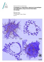
SIM imaging showed changes in the morphology of mitochondria and plasma membrane in approximately 50-60% of macrophages following LPS challenge (1μg/ml for 24 h), but no detectable changes in mitochondria or plasma membrane were observed after TNF-alpha (1ng/ml for 24 hr) or Con-A challenge (1μg/ml for 24 hr). QPM revealed that the phase value decreased by approximately 18% in LPS-challenged macrophages after 24 hr as compared to controls. In contrast, mitochondrial morphology appeared different in trophoblasts following TNF-alpha challenge under similar conditions. No effect was seen in trophoblast morphology following either LPS or Con-A challenge. Through SIM and QPM it was found that the effect on the morphology of macrophages can be detected following 2hr of incubation with LPS, whereas The Griess method was unable to detect increased amounts of NO before 4hr.
Our results suggest that the cells which are responsible for the mother-fetus crosstalk, especially macrophages and trophoblasts, respond differently to various inflammatory agents. Additionally, SIM and QPM are shown to be live-cell friendly, useful tools to evaluate sub-cellular mechanisms associated with inflammation mediated pregnancy complications. | en_US |
| dc.identifier.uri | https://hdl.handle.net/10037/11892 | |
| dc.language.iso | eng | en_US |
| dc.publisher | UiT Norges arktiske universitet | en_US |
| dc.publisher | UiT The Arctic University of Norway | en_US |
| dc.rights.accessRights | openAccess | en_US |
| dc.rights.holder | Copyright 2017 The Author(s) | |
| dc.rights.uri | https://creativecommons.org/licenses/by-nc-sa/3.0 | en_US |
| dc.rights | Attribution-NonCommercial-ShareAlike 3.0 Unported (CC BY-NC-SA 3.0) | en_US |
| dc.subject.courseID | FYS-3900 | |
| dc.subject | VDP::Mathematics and natural science: 400::Physics: 430 | en_US |
| dc.subject | VDP::Matematikk og Naturvitenskap: 400::Fysikk: 430 | en_US |
| dc.title | Investigation of inflammatory response of macrophages and trophoblasts using structured illumination microscopy | en_US |
| dc.type | Master thesis | en_US |
| dc.type | Mastergradsoppgave | en_US |


 English
 norsk
norsk
