| dc.contributor.author | Zhou, Wen | |
| dc.contributor.author | Reinstein, Dan Z. | |
| dc.contributor.author | Archer, Timothy J. | |
| dc.contributor.author | Chen, Xiangjun | |
| dc.contributor.author | Utheim, Tor Paaske | |
| dc.contributor.author | Feng, Yue | |
| dc.contributor.author | Stojanovic, Aleksandar | |
| dc.date.accessioned | 2022-03-23T09:58:06Z | |
| dc.date.available | 2022-03-23T09:58:06Z | |
| dc.date.issued | 2021-07-01 | |
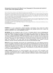
| dc.description.abstract | <p>PURPOSE: To assess intraoperative stromal topography measurements using swept-source optical coherence tomography (OCT)–based topography/tomography after epithelial removal and to analyze the epithelial contribution to the corneal topography and optics.
<p>METHODS: This was a prospective series of 22 eyes of 19 patients referred to receive phototherapeutic keratotomy (PTK) for treatment of recurrent corneal erosion and a control group of 22 virgin eyes. Swept-source OCT corneal topography/tomography was obtained immediately before and immediately after mechanical deepithelialization before PTK. Epithelial thickness maps were obtained before the surgery using spectral-domain OCT in the control group and as a reference in the group with anterior basement membrane dystrophy. Topographic and optical characteristics, including the curvature, astigmatism, asphericity, and higher order aberrations of the cornea before and after deepithelialization were compared, and their differences correlated with the measurements derived from the epithelial thickness maps.
<p>RESULTS: Stromal topography measurements after deepithelialization were easily obtained and showed excellent repeatability. Assessment of corneal edema induced by deepithelialization revealed that it did not significantly affect the measured parameters. The stromal surface was steeper by 1.28 diopters, had higher with-the-rule astigmatism by 0.41 diopters, was more prolate, and had more higher order aberrations compared to the intact epithelialized corneal surface. These differences correlated well with the parameters derived from epithelial thickness maps.
<p>CONCLUSIONS: Measurement of stromal topography using swept-source OCT immediately after mechanical deepithelialization may be a viable method in therapeutic refractive surgery, where stromal topography-guided ablation is needed. A significant epithelial contribution to anterior corneal topography and optics was confirmed. | en_US |
| dc.identifier.citation | Zhou, Reinstein, Archer, Chen, Utheim, Feng, Stojanovic. Intraoperative swept-source OCT–based corneal topography for measurement and analysis of stromal surface after epithelial removal. Journal of refractive surgery. 2021;37(7):484-492 | en_US |
| dc.identifier.cristinID | FRIDAID 2005237 | |
| dc.identifier.doi | 10.3928/1081597X-20210405-01 | |
| dc.identifier.issn | 1081-597X | |
| dc.identifier.issn | 1938-2391 | |
| dc.identifier.uri | https://hdl.handle.net/10037/24507 | |
| dc.language.iso | eng | en_US |
| dc.publisher | Slack | en_US |
| dc.relation.journal | Journal of refractive surgery | |
| dc.rights.accessRights | openAccess | en_US |
| dc.rights.holder | Copyright 2021 The Author(s) | en_US |
| dc.title | Intraoperative swept-source OCT–based corneal topography for measurement and analysis of stromal surface after epithelial removal | en_US |
| dc.type.version | acceptedVersion | en_US |
| dc.type | Journal article | en_US |
| dc.type | Tidsskriftartikkel | en_US |
| dc.type | Peer reviewed | en_US |


 English
 norsk
norsk