| dc.contributor.advisor | de Weerd, Louis | |
| dc.contributor.author | Weum, Sven | |
| dc.date.accessioned | 2016-09-16T07:07:24Z | |
| dc.date.available | 2016-09-16T07:07:24Z | |
| dc.date.issued | 2013-04-29 | |
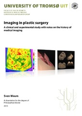
| dc.description.abstract | This thesis is based on four papers that have imaging techniques used in breast reconstruction as the common denominator. Each paper describes the results of a study. The purpose of the first three studies was to evaluate the use of dynamic infrared thermography (DIRT) as an imaging technique for perforator mapping in breast reconstruction with a deep inferior epigastric perforator (DIEP) flap. The purpose of the fourth study was to evaluate the form stability of the Style 410 anatomically shaped cohesive silicone gel-filled breast implant using magnetic resonance imaging (MRI). The studies reported cover a wide spectrum of imaging methods and all four illustrate how imaging may answer questions raised by the plastic surgeon. The first paper reports an experimental study in a university laboratory, the other three report clinical studies performed in a hospital setting.
DIRT was introduced at the University Hospital North Norway in 2002. It was observed that DIRT could be a promising method for perforator mapping in breast reconstructive surgery with a DIEP flap, but scientific evidence for such use of DIRT was lacking. The results from the first three studies provide scientific evidence to support the use of DIRT in the preoperative planning of DIEP flaps in autologous breast reconstruction. DIRT can replace computed tomographic angiography (CTA), which is today’s gold standard, as an imaging technique for preoperative perforator mapping. Such will have great advantages for patients. Unlike CTA, the non-invasive technique DIRT does not require exposure to ionizing radiation or the use of an intravenous contrast medium.
In the fourth study MRI is used in a novel way to visualize the behavior of the Style 410 breast implant in vivo as the body position is changed from supine to prone. The results show that the dimensions of the implant are influenced by the body position. The implant is therefore not form-stable with respect to its dimensions provided by the manufacturer, however, it nevertheless remains anatomically shaped with its largest projection in the lowest pole in both positions. Such knowledge on the behavior of the implant after implantation may help the surgeon in the preoperative planning and provides a better basis for patient information about the possible final result.
All four studies illustrate the value of interdisciplinary collaboration between the radiologist and plastic surgeon. The first three provide scientific support for the clinical use of DIRT as an imaging technique for perforator mapping while the fourth uses the well-established method MRI to answer questions that would otherwise be difficult to answer from the surgeon’s clinical point of view. | en_US |
| dc.description.doctoraltype | ph.d. | en_US |
| dc.description.popularabstract | Brystkreft rammer mer enn 2.500 norske kvinner årlig, og mange må fjerne brystet. Man kan nå lage nytt bryst av hud og fett fra magen, men slik kirurgi kan være risikofylt. For å unngå komplikasjoner må kirurgen ha oversikt over de små blodårene i bukveggen. I dag brukes CT rutinemessig for å kartlegge blodårene. En av ulempene med CT er at pasienten påføres en betydelig dose røntgenstråler. Sven Weum har forsket på bruk av varmekamera som et alternativ til CT og resultatene viser at varmekamera er like pålitelig som CT. Undersøkelsen er enkel og gir ingen skadelig stråling.
Silikonproteser kan også brukes til brystrekonstruksjon. Det er nå vanlig å bruke anatomisk formede silikonproteser da disse forventes å gi et bedre kosmetisk resultat enn tradisjonelle runde proteser. Men det har ikke tidligere vært gjort forskning som kartlegger protesenes form og oppførsel etter operasjonen. I avhandlingen brukes MR for å kartlegge protesenes dimensjoner i rygg- og mageleie. Resultatene viser at protesene har anatomisk form samtidig som de er myke nok til å forandre dimensjoner når kroppens stilling forandres. | en_US |
| dc.description.sponsorship | Universitetssykehuset Nord-Norge: D-stilling som lege i spesialisering og forskningsdager som overlege ved Røntgenavdelingen.
Helse Nord: Samskrivingsstipend 2012
Norwegian Research School in Medical Imaging: Travel and research grant | en_US |
| dc.description | The papers of this thesis are not available in Munin. <br>
Paper I : Miland, Å. O., de Weerd, L., Weum, S., Mercer, J. B.: “Visualising vascular perfusion in isolated human abdominal skin flaps using dynamic infrared thermography and indocyanine green fluorescence video angiography.” Available in <a href=http://dx.doi.org/10.1007/s00238-008-0280-9>European Journal of Plastic Surgery 2008, 31:235-42 </a> <br>
Paper II: de Weerd, L., Weum, S. Mercer, J. B.: “The value of dynamic infrared thermography (DIRT) in perforator selection and planning of DIEP flaps.” Available in <a href=http://dx.doi.org/10.1097/SAP.0b013e318190321e>Annals of Plastic Surgery 2009; 63(3):274-9 </a> <br>
Paper III: Weum, S., Mercer, J. B., de Weerd, L.: “Perforator mapping in breast reconstruction: A comparative study of dynamic infrared thermography (DIRT), computed tomographic angiography (CTA) and hand-‐held Doppler.” (Manuscript). Published version with title “Evaluation of dynamic infrared thermography as an alternative to CT angiography for perforator mapping in breast reconstruction: a clinical study” available in <a href=http://dx.doi.org/10.1186/s12880-016-0144-x> BMC Medical Imaging 2016, 16:43 </a> <br>
Paper IV: Weum S., de Weerd L, Kristiansen B.: “Form stability of Style 410 anatomically shape cohesive silicone gel-‐filled breast implant in subglandular breast augmentation evaluated with magnetic resonance imaging.” Available in <a href=http://dx.doi.org/10.1097/PRS.0b013e3181f95aba>Plastic and Reconstructive Surgery 2011, 127(1):409-13. </a> | en_US |
| dc.identifier.uri | https://hdl.handle.net/10037/9697 | |
| dc.identifier.urn | URN:NBN:no-uit_munin_9214 | |
| dc.language.iso | eng | en_US |
| dc.rights.accessRights | openAccess | |
| dc.rights.holder | Copyright 2013 The Author(s) | |
| dc.rights.uri | https://creativecommons.org/licenses/by-nc-sa/3.0 | en_US |
| dc.rights | Attribution-NonCommercial-ShareAlike 3.0 Unported (CC BY-NC-SA 3.0) | en_US |
| dc.subject | VDP::Medisinske Fag: 700::Klinisk medisinske fag: 750::Radiologi og bildediagnostikk: 763 | en_US |
| dc.subject | VDP::Medical disciplines: 700::Clinical medical disciplines: 750::Radiology and diagnostic imaging: 763 | en_US |
| dc.subject | VDP::Medisinske Fag: 700::Klinisk medisinske fag: 750::Plastisk kirurgi: 785 | en_US |
| dc.subject | VDP::Medical disciplines: 700::Clinical medical disciplines: 750::Plastic surgery: 785 | en_US |
| dc.subject | VDP::Medisinske Fag: 700::Basale medisinske, odontologiske og veterinærmedisinske fag: 710::Human og veterinærmedisinsk fysiologi: 718 | en_US |
| dc.subject | VDP::Medical disciplines: 700::Basic medical, dental and veterinary science disciplines: 710::Human and veterinary science physiology: 718 | en_US |
| dc.title | Imaging in plastic surgery. A clinical and experimental study with notes on the history of medical imaging. | en_US |
| dc.type | Doctoral thesis | en_US |
| dc.type | Doktorgradsavhandling | en_US |


 English
 norsk
norsk


