English
 norsk
norskBrowsing by Author "Agarwal, Krishna"
Now showing items 21-39 of 39
-
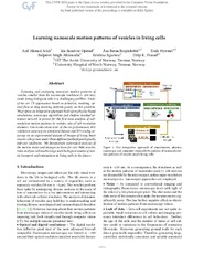
Learning nanoscale motion patterns of vesicles in living cells
(Chapter; Bokkapittel, 2020)Detecting and analyzing nanoscale motion patterns of vesicles, smaller than the microscope resolution (~250 nm), inside living biological cells is a challenging problem. State-of-the-art CV approaches based on detection, tracking, optical flow or deep learning perform poorly for this problem. We propose an integrative approach, built upon physics based simulations, nanoscopy algorithms, and shallow ... -
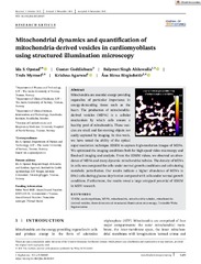
Mitochondrial dynamics and quantification of mitochondria-derived vesicles in cardiomyoblasts using structured illumination microscopy
(Journal article; Tidsskriftartikkel; Peer reviewed, 2021-11-12)Mitochondria are essential energy-providing organelles of particular importance in energy-demanding tissue such as the heart. The production of mitochondria-derived vesicles (MDVs) is a cellular mechanism by which cells ensure a healthy pool of mitochondria. These vesicles are small and fast-moving objects not easily captured by imaging. In this work, we have tested the ability of the optical ... -
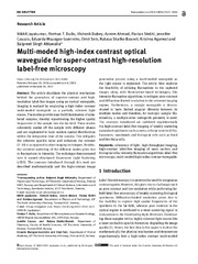
Multi-moded high-index contrast optical waveguide for super-contrast high-resolution label-free microscopy
(Journal article; Tidsskriftartikkel; Peer reviewed, 2022-06-20)The article elucidates the physical mechanism behind the generation of superior-contrast and highresolution label-free images using an optical waveguide. Imaging is realized by employing a high index contrast multi-moded waveguide as a partially coherent light source. The modes provide near-field illumination of unlabeled samples, thereby repositioning the higher spatial frequencies of the ... -
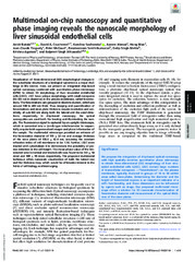
Multimodal on-chip nanoscopy and quantitative phase imaging reveals the nanoscale morphology of liver sinusoidal endothelial cells
(Journal article; Tidsskriftartikkel; Peer reviewed, 2021-11-23)Visualization of three-dimensional (3D) morphological changes in the subcellular structures of a biological specimen is a major challenge in life science. Here, we present an integrated chip-based optical nanoscopy combined with quantitative phase microscopy (QPM) to obtain 3D morphology of liver sinusoidal endothelial cells (LSEC). LSEC have unique morphology with small nanopores (50-300 nm in ... -
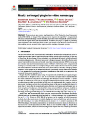
MusiJ: an ImageJ plugin for video nanoscopy
(Journal article; Tidsskriftartikkel; Peer reviewed, 2020-04-14)We present an open-source implementation of the fluctuation-based nanoscopy method MUSICAL for ImageJ. This implementation improves the algorithm’s computational efficiency and takes advantage of multi-threading to provide orders of magnitude faster reconstructions than the original MATLAB implementation. In addition, the plugin is capable of generating super-resolution videos from large stacks of ... -
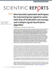
Non-heuristic automatic techniques for overcoming low signal-to-noise-ratio bias of localization microscopy and multiple signal classification algorithm
(Journal article; Tidsskriftartikkel; Peer reviewed, 2018-03-21)Localization microscopy and multiple signal classification algorithm use temporal stack of image frames of sparse emissions from fluorophores to provide super-resolution images. Localization microscopy localizes emissions in each image independently and later collates the localizations in all the frames, giving same weight to each frame irrespective of its signal-to-noise ratio. This results in a ... -
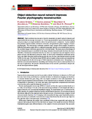
Object detection neural network improves Fourier ptychography reconstruction
(Journal article; Tidsskriftartikkel; Peer reviewed, 2020-11-23)High resolution microscopy is heavily dependent on superb optical elements and superresolution microscopy even more so. Correcting unavoidable optical aberrations during post-processing is an elegant method to reduce the optical system’s complexity. A prime method that promises superresolution, aberration correction, and quantitative phase imaging is Fourier ptychography. This microscopy technique ... -
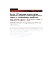
On-chip TIRF nanoscopy by applying Haar wavelet kernel analysis on intensity fluctuations induced by chip illumination
(Journal article; Tidsskriftartikkel; Peer reviewed, 2020-11-09)Photonic-chip based TIRF illumination has been used to demonstrate several on-chip optical nanoscopy methods. The sample is illuminated by the evanescent field generated by the electromagnetic wave modes guided inside the optical waveguide. In addition to the photokinetics of the fluorophores, the waveguide modes can be further exploited for introducing controlled intensity fluctuations for exploitation ... -
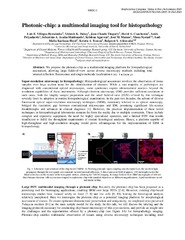
Photonic-chip: a multimodal imaging tool for histopathology
(Conference object; Konferansebidrag, 2021-04)We propose the photonic-chip as a multimodal imaging platform for histopathological assessment, allowing large fields-of-view across diverse microscopy methods including total internal reflection fluorescence and single-molecule localization. -
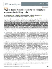
Physics-based machine learning for subcellular segmentation in living cells
(Journal article; Tidsskriftartikkel; Peer reviewed, 2021-12-15)Segmenting subcellular structures in living cells from fluorescence microscope images is a ground truth (GT)-deficient problem. The microscopes’ three-dimensional blurring function, finite optical resolution due to light diffraction, finite pixel resolution and the complex morphological manifestations of the structures all contribute to GT-hardness. Unsupervised segmentation approaches are quite ... -
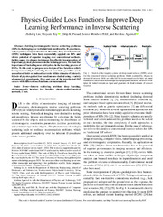
Physics-Guided Loss Functions Improve Deep Learning Performance in Inverse Scattering
(Journal article; Tidsskriftartikkel; Peer reviewed, 2022-03-15)Solving electromagnetic inverse scattering problems (ISPs) is challenging due to the intrinsic nonlinearity, ill-posedness, and expensive computational cost. Recently, deep neural network (DNN) techniques have been successfully applied on ISPs and shown potential of superior imaging over conventional methods. In this paper, we discuss techniques for effective incorporation of important physical ... -
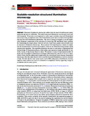
Scalable-resolution structured illumination microscopy
(Journal article; Tidsskriftartikkel; Peer reviewed, 2022-11-15)Structured illumination microscopy suffers from the need of sophisticated instrumentation and precise calibration. This makes structured illumination microscopes costly and skill-dependent. We present a novel approach to realize super-resolution structured illumination microscopy using an alignment non-critical illumination system and a reconstruction algorithm that does not need illumination ... -
Silicon substrate significantly alters dipole-dipole resolution in coherent microscope
(Journal article; Tidsskriftartikkel; Peer reviewed, 2020-12-16)Considering a coherent microscopy setup, influences of the substrate below the sample in the imaging performances are studied, with a focus on high refractive index substrate such as silicon. Analytical expression of 3D full-wave vectorial point spread function, i.e. the dyadic Green's function is derived for the optical setup together with the substrate. Numerical analysis are performed in order ... -
Single-shot multispectral quantitative phase imaging of biological samples using deep learning
(Journal article; Tidsskriftartikkel; Peer reviewed, 2023-05-16)Multispectral quantitative phase imaging (MS-QPI) is a high-contrast label-free technique for morphological imaging of the specimens. The aim of the present study is to extract spectral dependent quantitative information in single-shot using a highly spatially sensitive digital holographic microscope assisted by a deep neural network. There are three different wavelengths used in our method: 𝜆=532 , ... -
Soft thresholding schemes for multiple signal classification algorithm
(Journal article; Tidsskriftartikkel; Peer reviewed, 2020-10-28)Multiple signal classification algorithm (MUSICAL) exploits temporal fluctuations in fluorescence intensity to perform super-resolution microscopy by computing the value of a super-resolving indicator function across a fine sample grid. A key step in the algorithm is the separation of the measurements into signal and noise subspaces, based on a single user-specified parameter called the threshold. ... -
Three-dimensional structured illumination microscopy data of mitochondria and lysosomes in cardiomyoblasts under normal and galactose-adapted conditions
(Journal article; Tidsskriftartikkel; Peer reviewed, 2022-03-23)This three-dimensional structured illumination microscopy (3DSIM) dataset was generated to highlight the suitability of 3DSIM to investigate mitochondria-derived vesicles (MDVs) in H9c2 cardiomyoblasts in living or fxed cells. MDVs act as a mitochondria quality control mechanism. The cells were stably expressing the tandem-tag eGFP-mCherry-OMP25-TM (outer mitochondrial membrane) which can be used ... -
Viewing life without labels under optical microscopes
(Journal article; Tidsskriftartikkel; Peer reviewed, 2023-05-25)Optical microscopes today have pushed the limits of speed, quality, and observable space in biological specimens revolutionizing how we view life today. Further, specific labeling of samples for imaging has provided insight into how life functions. This enabled label-based microscopy to percolate and integrate into mainstream life science research. However, the use of labelfree microscopy has been ... -
Virtual labeling of mitochondria in living cells using correlative imaging and physics-guided deep learning
(Journal article; Tidsskriftartikkel; Peer reviewed, 2022-09-28)Mitochondria play a crucial role in cellular metabolism. This paper presents a novel method to visualize mitochondria in living cells without the use of fluorescent markers. We propose a physics-guided deep learning approach for obtaining virtually labeled micrographs of mitochondria from bright-field images. We integrate a microscope’s point spread function in the learning of an adversarial neural ...