English
 norsk
norskBlar i forfatter "Opstad, Ida Sundvor"
Viser treff 21-23 av 23
-
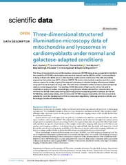
Three-dimensional structured illumination microscopy data of mitochondria and lysosomes in cardiomyoblasts under normal and galactose-adapted conditions
(Journal article; Tidsskriftartikkel; Peer reviewed, 2022-03-23)This three-dimensional structured illumination microscopy (3DSIM) dataset was generated to highlight the suitability of 3DSIM to investigate mitochondria-derived vesicles (MDVs) in H9c2 cardiomyoblasts in living or fxed cells. MDVs act as a mitochondria quality control mechanism. The cells were stably expressing the tandem-tag eGFP-mCherry-OMP25-TM (outer mitochondrial membrane) which can be used ... -
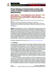
Virtual labeling of mitochondria in living cells using correlative imaging and physics-guided deep learning
(Journal article; Tidsskriftartikkel; Peer reviewed, 2022-09-28)Mitochondria play a crucial role in cellular metabolism. This paper presents a novel method to visualize mitochondria in living cells without the use of fluorescent markers. We propose a physics-guided deep learning approach for obtaining virtually labeled micrographs of mitochondria from bright-field images. We integrate a microscope’s point spread function in the learning of an adversarial neural ... -
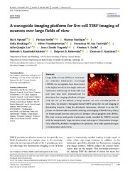
A waveguide imaging platform for live-cell TIRF imaging of neurons over large fields of view
(Journal article; Tidsskriftartikkel; Peer reviewed, 2020-02-17)Large fields of view (FOVs) in total internal reflection fluorescence microscopy (TIRFM) via waveguides have been shown to be highly beneficial for single molecule localisation microscopy on fixed cells [1,2] and have also been demonstrated for short‐term live‐imaging of robust cell types [3‐5], but not yet for delicate primary neurons nor over extended periods of time. Here, we present a waveguide‐based ...