English
 norsk
norskBlar i forfatter "Ströhl, Florian"
Viser treff 21-23 av 23
-
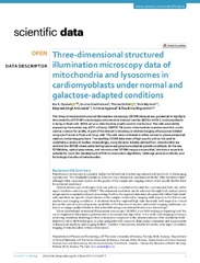
Three-dimensional structured illumination microscopy data of mitochondria and lysosomes in cardiomyoblasts under normal and galactose-adapted conditions
(Journal article; Tidsskriftartikkel; Peer reviewed, 2022-03-23)This three-dimensional structured illumination microscopy (3DSIM) dataset was generated to highlight the suitability of 3DSIM to investigate mitochondria-derived vesicles (MDVs) in H9c2 cardiomyoblasts in living or fxed cells. MDVs act as a mitochondria quality control mechanism. The cells were stably expressing the tandem-tag eGFP-mCherry-OMP25-TM (outer mitochondrial membrane) which can be used ... -
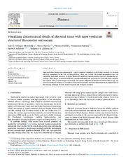
Visualizing ultrastructural details of placental tissue with super-resolution structured illumination microscopy
(Journal article; Tidsskriftartikkel; Peer reviewed, 2020-06-14)Super-resolution fluorescence microscopy is a widely employed technique in cell biology research, yet remains relatively unexplored in the field of histopathology. Here, we describe the sample preparation steps and acquisition parameters necessary to obtain fluorescent multicolor super-resolution structured illumination microscopy (SIM) images of both formalin-fixed paraffin-embedded and cryo-preserved ... -
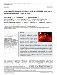
A waveguide imaging platform for live-cell TIRF imaging of neurons over large fields of view
(Journal article; Tidsskriftartikkel; Peer reviewed, 2020-02-17)Large fields of view (FOVs) in total internal reflection fluorescence microscopy (TIRFM) via waveguides have been shown to be highly beneficial for single molecule localisation microscopy on fixed cells [1,2] and have also been demonstrated for short‐term live‐imaging of robust cell types [3‐5], but not yet for delicate primary neurons nor over extended periods of time. Here, we present a waveguide‐based ...