

 English
 norsk
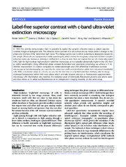
norskLabel-free superior contrast with c-band ultra-violet extinction microscopy
| dc.contributor.author | Wolfson, Deanna | |
| dc.contributor.author | Opstad, Ida Sundvor | |
| dc.contributor.author | Hansen, Daniel Henry | |
| dc.contributor.author | Mao, Hong | |
| dc.contributor.author | Ahluwalia, Balpreet Singh | |
| dc.contributor.author | Ströhl, Florian | |
| dc.date.accessioned | 2023-04-24T07:57:53Z | |
| dc.date.available | 2023-04-24T07:57:53Z | |
| dc.date.issued | 2023-03-03 | |
| dc.description.abstract | In 1934, Frits Zernike demonstrated that it is possible to exploit the sample’s refractive index to obtain superior contrast images of biological cells. The refractive index contrast of a cell surrounded by media yields a change in the phase and intensity of the transmitted light wave. This change can be due to either scattering or absorption caused by the sample. Most cells are transparent at visible wavelengths, which means the imaginary component of their complex refractive index, also known as extinction coefficient k, is close to zero. Here, we explore the use of c-band ultra-violet (UVC) light for high-contrast high-resolution label-free microscopy, as k is naturally substantially higher in the UVC than at visible wavelengths. Using differential phase contrast illumination and associated processing, we achieve a 7- to 300-fold improvement in contrast compared to visible-wavelength and UVA differential interference contrast microscopy or holotomography, and quantify the extinction coefficient distribution within liver sinusoidal endothelial cells. With a resolution down to 215 nm, we are, for the first time in a far-field label-free method, able to image individual fenestrations within their sieve plates which normally requires electron or fluorescence superresolution microscopy. UVC illumination also matches the excitation peak of intrinsically fluorescent proteins and amino acids and thus allows us to utilize autofluorescence as an independent imaging modality on the same setup. | en_US |
| dc.description.sponsorship | EU | en_US |
| dc.identifier.citation | Wolfson, Opstad, Hansen, Mao, Ahluwalia. Label-free superior contrast with c-band ultra-violet extinction microscopy. Light: Science & Applications (LSA). 2023 | en_US |
| dc.identifier.cristinID | FRIDAID 2131003 | |
| dc.identifier.doi | 10.1038/s41377-023-01105-6 | |
| dc.identifier.issn | 2095-5545 | |
| dc.identifier.issn | 2047-7538 | |
| dc.identifier.uri | https://hdl.handle.net/10037/29032 | |
| dc.language.iso | eng | en_US |
| dc.publisher | Springer Nature | en_US |
| dc.relation.journal | Light: Science & Applications (LSA) | |
| dc.relation.projectID | info:eu-repo/grantAgreement/ERC/H2020/ 836355/EU/Development of Deep-UV Quantitative Microscopy for the Study of Mitochondrial Dysfunction/MitoQuant/ | en_US |
| dc.rights.accessRights | openAccess | en_US |
| dc.rights.holder | Copyright 2023 The Author(s) | en_US |
| dc.rights.uri | https://creativecommons.org/licenses/by/4.0 | en_US |
| dc.rights | Attribution 4.0 International (CC BY 4.0) | en_US |
| dc.title | Label-free superior contrast with c-band ultra-violet extinction microscopy | en_US |
| dc.type.version | publishedVersion | en_US |
| dc.type | Journal article | en_US |
| dc.type | Tidsskriftartikkel | en_US |
| dc.type | Peer reviewed | en_US |
Tilhørende fil(er)
Denne innførselen finnes i følgende samling(er)

Med mindre det står noe annet, er denne innførselens lisens beskrevet som Attribution 4.0 International (CC BY 4.0)