| dc.contributor.author | Wolfson, Deanna | |
| dc.contributor.author | Villegas, Luis | |
| dc.contributor.author | Opstad, Ida Sundvor | |
| dc.contributor.author | Birgisdottir, Åsa birna | |
| dc.contributor.author | Øie, Cristina Ionica | |
| dc.contributor.author | Ahluwalia, Balpreet Singh | |
| dc.date.accessioned | 2023-10-06T06:00:58Z | |
| dc.date.available | 2023-10-06T06:00:58Z | |
| dc.date.issued | 2018 | |
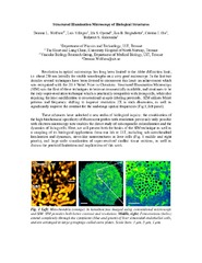
| dc.description.abstract | <p>Resolution in optical microscopy has long been limited to the Abbe diffraction limit, i.e. about 250 nm laterally for visible wavelengths on a very good microscope. In the last two decades several techniques have been devised to circumvent this limit: an achievement which was recognized with the 2014 Nobel Prize in Chemistry. Structured Illumination Microscopy (SIM) was the first of these techniques to become commercially available, and continues to be the only super-resolution technique which is practically compatible with living cells, while also requiring the least modification to conventional sample-labeling protocols. SIM utilizes Moiré patterns and frequency shifting to improve resolution 2X in each dimension, as well as significantly improve the contrast for the mid-range spatial frequencies.
<p>These advances have unlocked a new realm of biological inquiry: the combination of the high biochemical specificity of fluorescent probes with resolution previously only possible with electron microscopy now enables the direct study of sub-organelle colocalization and the dynamics of living cells. Here, we will present both the basics of the SIM technique as well as a sampling of its biological applications from our lab at UiT, including sub-mitochondrial localization and dynamics, sieve-like nanostructures in liver cells, and large-scale visualization of super-resolved cardiac tissue sections, as well as discuss the practical limitations and implications of this work. | en_US |
| dc.description | Abstract of presentation held at Norwegian Electro-Optics Meeting, Henningsvær, Norway, 2-4 May 2018. | en_US |
| dc.identifier.cristinID | FRIDAID 1655112 | |
| dc.identifier.uri | https://hdl.handle.net/10037/31475 | |
| dc.language.iso | eng | en_US |
| dc.rights.accessRights | openAccess | en_US |
| dc.title | Structured Illumination Microscopy of Biological Structures | en_US |
| dc.type | Conference object | en_US |
| dc.type | Konferansebidrag | en_US |


 English
 norsk
norsk