English
 norsk
norskBlar i forfatter Det helsevitenskapelige fakultet "Huser, Thomas Rolf"
Viser treff 1-3 av 3
-
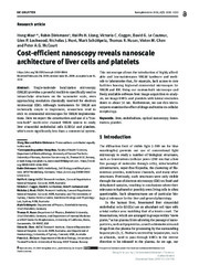
Cost-efficient nanoscopy reveals nanoscale architecture of liver cells and platelets
(Journal article; Tidsskriftartikkel; Peer reviewed, 2019-07-09)Single-molecule localization microscopy (SMLM) provides a powerful toolkit to specifically resolve intracellular structures on the nanometer scale, even approaching resolution classically reserved for electron microscopy (EM). Although instruments for SMLM are technically simple to implement, researchers tend to stick to commercial microscopes for SMLM implementations. Here we report the construction ... -
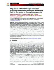
High-speed TIRF and 2D super-resolution structured illumination microscopy with a large field of view based on fiber optic components
(Journal article; Tidsskriftartikkel; Peer reviewed, 2023-08-16)Super-resolved structured illumination microscopy (SR-SIM) is among the most flexible, fast, and least perturbing fluorescence microscopy techniques capable of surpassing the optical diffraction limit. Current custom-built instruments are easily able to deliver two-fold resolution enhancement at video-rate frame rates, but the cost of the instruments is still relatively high, and the physical size ... -
Multimodal super-resolution optical microscopy visualizes the close connection between membrane and the cytoskeleton in liver sinusoidal endothelial cell fenestrations
(Journal article; Tidsskriftartikkel; Peer reviewed, 2015)Liver sinusoidal endothelial cells (LSECs) act as a filter between blood and the hepatocytes. LSECs are highly fenestrated cells; they contain transcellular pores with diameters between 50 to 200 nm. The small sizes of the fenestrae have so far prohibited any functional analysis with standard and advanced light microscopy techniques. Only the advent of super-resolution optical fluorescence microscopy ...