| dc.contributor.advisor | McCourt, Peter | |
| dc.contributor.author | Mao, Hong | |
| dc.date.accessioned | 2021-01-05T09:36:56Z | |
| dc.date.available | 2021-01-05T09:36:56Z | |
| dc.date.issued | 2021-01-22 | |
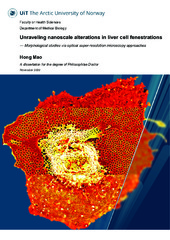
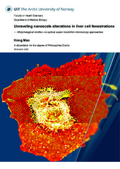
| dc.description.abstract | The endothelium makes up the innermost cell layer of blood vessels. It consists of a thin layer of simple squamous cells, forming an interface between circulating blood and the surrounding tissue. Endothelial cells of different vascular beds are specialized according to tissue-specific functions. For this project emphasis was placed upon high-resolution methods enabling the study of liver sinusoidal endothelial cells (LSECs) below the diffraction limit of visible light (~200 nm). LSECs have unusual morphology with as much of 20% of their surface covered with cellular fenestrations - holes through the cells of 50-300 nm diameter. These allow bi-directional flow of plasma from the sinusoids to the surrounding hepatocytes, while retaining blood cells in the sinusoidal lumen. Little is known about the function of fenestrations, their regulation, and their role in the transfer of metabolites, viruses, lipoproteins and pharmaceuticals to other cells of the liver.
There are two major challenges with the study of LSEC fenestrations; i) the majority have diameters smaller than the diffraction limit of visible light and; ii) they disappear rapidly in cultured LSEC, and there are no cell line alternatives that express fenestrations. To address the first challenge, the project used classical super resolution imaging technologies such as scanning electron microscopy, and two novel super-resolution optical microscopy modalities: dSTORM (direct stochastic optical reconstruction microscopy) and SIM (structured illumination microscopy) to study the in vitro effects of xanthines, sildenafil and oxidized LDL on LSEC fenestrations. One of the xanthines, theobromine, and sildenafil increased both the frequency and diameter of fenestrations in cultured LSEC. While oxidized LDL caused major disruptions in LSEC fenestration morphology. Finally, to address the second challenge, namely the rapid loss of fenestrations in LSEC, a cryopreservation method for freshly isolated LSEC was developed such that they can be used at researchers’ convenience, rather than directly after isolation from live | en_US |
| dc.description.doctoraltype | ph.d. | en_US |
| dc.description.popularabstract | Our livers are the largest organ in our bodies, and every minute 30% of our blood flows through them. Our livers have two main purposes. The first is to supply the blood (and thus all of the body’s other organs) with essential proteins, hormones, glucose (between meals) and lipoproteins such as low-density lipoprotein (LDL): cholesterol. The second is to clean the blood by removing old or damaged proteins, medicines, excess glucose and excess cholesterol. Much of this supply to - and removal from - the blood goes via small holes (“fenestrations”) in liver sinusoidal endothelial cells (LSECs). As much as 20% of the LSEC surface is perforated with these fenestrations, and LSEC lines the all blood vessels permeating through the liver. In addition, if all the LSEC from a human liver were laid out flat, they would cover the area of one tennis court. This makes LSEC, with their fenestrations, the largest “filter-cell” system in the body.
These fenestrations are really small, and range from 50 to 300 nanometers in diameter in normal healthy (and young) people. However, when we age or have liver disease, the fenestrations become fewer and smaller. This restricts the filtration of material to and from the liver, and this will have consequences for the entire body. For example, medicines are removed from the blood by the liver through fenestrations in a process called detoxification. Young people with healthy livers easily detoxify medicines after the medicines have done their job. However, in older people, detoxification goes more slowly because they have smaller and fewer fenestrations, so medicine dosages safe for young people can be poisonous for older people.
The purpose of this thesis is to develop novel ways to look at fenestrations in LSEC via innovative microscope technologies, and find ways to keep fenestrations as we age. Until recently it has been impossible to study LSEC fenestrations with ordinary light microscopes because fenestrations are so small. However, we now have new microscopy “tricks” to see fenestrations in living LSEC, and study how they behave and respond to different treatments in real-time. We have improved on these “tricks” by building our super-resolution (dSTORM: direct stochastic optical reconstruction microscopy) microscope for 10% of the cost of a commercial dSTORM system, and used it to study LSEC fenestrations. We published this study with a detailed set of building instructions for the dSTORM microscope that other researchers can use. Using our “home-built” dSTORM, and a new commercial structured illumination microscope (SIM), we studied the effect of various compounds on LSEC fenestrations. These compounds included xanthines (caffeine, theobromine, theophylline and others), medicines such as sildenafil, and oxidized LDL (oxLDL): cholesterol. We found that theobromine and sildenafil increased the size and number of fenestrations on LSEC. We found that oxLDL caused major disruptions in LSEC fenestrations – this compound is found in large amounts in patients with severe heart disease, and is clearly damaging for LSEC.
In summary, we customized a cost-efficient microscope to visualize very small filter holes (fenestrations) in liver cells. These holes are crucial for good health, and our studies are assisting us to lead ways to maintain these functional fenestrations during aging and liver disease. These methods have helped to show that theobromine (from dark chocolate) and sildenafil (a.k.a Viagra) appear to be beneficial for LSEC fenestrations in in vitro experiments. These findings are still to be confirmed in vivo in animal models. We also showed that oxidized LDL has very negative in vitro effects on LSEC fenestrations. This would suggest that consuming anti-oxidants would prevent the formation of oxidized LDL and would contribute to better liver health. | en_US |
| dc.description.sponsorship | Grants from the Tromsø Research Foundation/Trond Mohn, the University of Tromsø-The Arctic University of Norway, the Research Council of Norway FRIMED grant no. 262538, and Marie Sklodowska-Curie Grant Agreement No. 766181, project: DeLIVER. | en_US |
| dc.identifier.uri | https://hdl.handle.net/10037/20170 | |
| dc.language.iso | eng | en_US |
| dc.publisher | UiT The Arctic University of Norway | en_US |
| dc.publisher | UiT Norges arktiske universitet | en_US |
| dc.relation.haspart | <p>Paper I: Mao, H., Diekmann, R., Liang, H.P.H., Cogger, V.C., Le Couteur, D.G., Lockwood, G.P., ... McCourt, P.A.G. (2091). Cost-efficient nanoscopy reveals nanoscale architecture of liver cells and platelets. <i>Nanophotonics, 8</i>(7), 1299-1313. Also available in Munin at <a href=https://hdl.handle.net/10037/16255>https://hdl.handle.net/10037/16255</a>.
<p>Paper II: Mao, H., Szafrańska, K., Wolfson, D.L., Ahluwalia, B.S., Whitechurch, C.B., Lockwood, G.P., … McCourt, P.A.G. Effect of caffeine, theobromine and other xanthines on liver sinusoidal endothelial cell ultrastructure. (Manuscript). Available in the file thesis_entire.pdf.
<p>Paper III: Mao, H., Kruse, L.D., Li, R., Oteiza, A., Cogger, V.C., Le Couteur, D., … McCourt, P.A.G. Impact of oxidized low-density protein on liver sinusoidal endothelial cells ultrastructure. (Manuscript). Available in the file thesis_entire.pdf.
<p>Paper IV: Mönkemöller, V., Mao, H., Hübner, W., Dumitriu, G., Heimann, P., Levy, G., … Øie, C. (2018). Primary rat LSECs preserve their characteristic phenotype after cryopreservation. <i>Scientific Reports, 8</i>, 14657. Also available in Munin at <a href=https://hdl.handle.net/10037/13983>https://hdl.handle.net/10037/13983</a>. | en_US |
| dc.relation.projectID | info:eu-repo/grantAgreement/RCN/FRIMED2/262538/Norway/Role of scavenger endothelial cells in elimination of virus// | en_US |
| dc.relation.projectID | info:eu-repo/grantAgreement/EC/H2020/766181/EU/Super-resolution optical microscopy of nanosized pore dynamics in endothelial cells/DeLIVER/ | en_US |
| dc.rights.accessRights | openAccess | en_US |
| dc.rights.holder | Copyright 2021 The Author(s) | |
| dc.rights.uri | https://creativecommons.org/licenses/by-nc-sa/4.0 | en_US |
| dc.rights | Attribution-NonCommercial-ShareAlike 4.0 International (CC BY-NC-SA 4.0) | en_US |
| dc.subject | VDP::Medical disciplines: 700::Basic medical, dental and veterinary science disciplines: 710 | en_US |
| dc.subject | VDP::Medisinske Fag: 700::Basale medisinske, odontologiske og veterinærmedisinske fag: 710 | en_US |
| dc.subject | VDP::Technology: 500::Nanotechnology: 630 | en_US |
| dc.subject | VDP::Teknologi: 500::Nanoteknologi: 630 | en_US |
| dc.subject | VDP::Teknologi: 500::Bioteknologi: 590 | en_US |
| dc.subject | VDP::Medical disciplines: 700::Health sciences: 800::Other health science disciplines: 829 | en_US |
| dc.subject | VDP::Medisinske Fag: 700::Helsefag: 800::Andre helsefag: 829 | en_US |
| dc.title | Unraveling nanoscale alterations in liver cell fenestrations - Morphological studies via optical super-resolution microscopy approaches | en_US |
| dc.type | Doctoral thesis | en_US |
| dc.type | Doktorgradsavhandling | en_US |


 English
 norsk
norsk

