

 English
 norsk
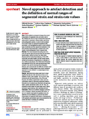
norskNovel approach to artefact detection and the definition of normal ranges of segmental strain and strain-rate values
| dc.contributor.author | Kornev, Mikhail | |
| dc.contributor.author | Caglayan, Hatice Aklay | |
| dc.contributor.author | Kudryavtsev, Alexander V | |
| dc.contributor.author | Malyutina, Sofia | |
| dc.contributor.author | Ryabikov, Andrew | |
| dc.contributor.author | Stylidis, Michael | |
| dc.contributor.author | Schirmer, Henrik | |
| dc.contributor.author | Rosner, Assami | |
| dc.date.accessioned | 2023-02-03T08:42:08Z | |
| dc.date.available | 2023-02-03T08:42:08Z | |
| dc.date.issued | 2022-12-13 | |
| dc.description.abstract | Aims - Strain artefacts are known to hamper the correct interpretation of segmental strain and strain-rate (S/SR). Defining the normal ranges of myocardial segmental deformation is important in clinical studies and routine echocardiographic practice. In order to define artefact-free normal ranges for segmental longitudinal S/SR parameters, we investigated the extent to which different types of artefacts and their segmental localisation in the three different myocardial layers created a bias in the results of echocardiographic strain measurements.<p> <p>Methods - The study included echocardiograms from men and women aged 40–69 years from two population-based studies, namely the Know Your Heart study (Russia) and the Tromsø Study (Norway). Of the 2207 individuals from these studies, 840 had normal results, defined as the absence of hypertension or indicators of any cardiovascular disease. Two-dimensional (2D) global and segmental S/SR of the three myocardial layers were analysed using speckle tracking echocardiography. Artefacts were assessed with two different methods: visual identification of image-artefacts and a novel conceptual approach of ‘curve-artefacts’ or unphysiological strain-curve formation.<p> <p>Results - Segmental strain values were found to have significantly reduced in the presence of strain-curve artefacts (14.9%±5.8% towards −20.7%±4.9%), and increased with the foreshortening of the 2D image. However, the individual global strain values were not substantially altered by discarding segmental artefacts. Reduction due to artefacts was observed in all segments, layers, systolic and diastolic strain, and SR. Thus, we presented normal ranges for basal-septal, basal, medial and apical segment groups after excluding artefacts.<p> <p>Conclusion - Strain-curve artefacts introduce systematic errors, resulting in reduced segmental S/SR values. In terms of artefact-robust global longitudinal strain, the detection of curve-artefacts is crucial for the correct interpretation of segmental S/SR patterns. Intersegmental S/SR gradients and artefacts need to be considered for the correct definition of normalcy and pathology. | en_US |
| dc.identifier.citation | Kornev M, Caglayan, Kudryavtsev AV, Malyutina S, Ryabikov A, Stylidis S, Schirmer H, Rosner A. Novel approach to artefact detection and the definition of normal ranges of segmental strain and strain-rate values. Open heart. 2022;9(2) | |
| dc.identifier.cristinID | FRIDAID 2121832 | |
| dc.identifier.doi | 10.1136/openhrt-2022-002136 | |
| dc.identifier.issn | 2053-3624 | |
| dc.identifier.uri | https://hdl.handle.net/10037/28483 | |
| dc.language.iso | eng | en_US |
| dc.publisher | BMJ Publishing Group | en_US |
| dc.relation.journal | Open heart | |
| dc.rights.holder | Copyright 2022 The Author(s) | en_US |
| dc.rights.uri | https://creativecommons.org/licenses/by-nc/4.0 | en_US |
| dc.rights | Attribution-NonCommercial 4.0 International (CC BY-NC 4.0) | en_US |
| dc.title | Novel approach to artefact detection and the definition of normal ranges of segmental strain and strain-rate values | en_US |
| dc.type.version | publishedVersion | en_US |
| dc.type | Journal article | en_US |
| dc.type | Tidsskriftartikkel | en_US |
| dc.type | Peer reviewed | en_US |
Tilhørende fil(er)
Denne innførselen finnes i følgende samling(er)

Med mindre det står noe annet, er denne innførselens lisens beskrevet som Attribution-NonCommercial 4.0 International (CC BY-NC 4.0)