

 English
 norsk
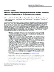
norskObserver agreement of imaging measurements used for evaluation of dentofacial deformity in juvenile idiopathic arthritis
| dc.contributor.author | Fischer, Johannes Maria | |
| dc.contributor.author | Halbig, Josefine Mareile | |
| dc.contributor.author | Augdal, Thomas Angell | |
| dc.contributor.author | Angenete, Oskar W | |
| dc.contributor.author | Stoustrup, Peter | |
| dc.contributor.author | Kristensen, Kasper Dahl | |
| dc.contributor.author | Skeie, Marit Slåttelid | |
| dc.contributor.author | Tylleskär, Karin | |
| dc.contributor.author | Rosén, Annika | |
| dc.contributor.author | Shi, Xie-Qi | |
| dc.contributor.author | Rosendahl, Karen | |
| dc.date.accessioned | 2023-02-08T14:16:47Z | |
| dc.date.available | 2023-02-08T14:16:47Z | |
| dc.date.issued | 2022-08-02 | |
| dc.description.abstract | Objectives: To examine the precision of imaging measures commonly used to assess mandibular morphology in children and adolescents with juvenile idiopathic arthritis (JIA). Secondly, to compare cone-beam computed tomography (CBCT) and magnetic resonance imaging (MRI) in the measurement of condylar height.<p> <p>Methods: Those included were children diagnosed with JIA during 2015–18 who had had an MRI, a CBCT of the temporomandibular joints (TMJs) and a lateral cephalogram (ceph) of the head within one month of each other. Agreement within and between observers and methods was examined using Bland-Altman mean-difference plots and 95% limits of agreement (LOA). A 95% LOA within 15% of the sample mean was considered acceptable. Minimal detectable change (MDC) within and between observers was estimated.<p> <p>Results: 90 patients (33 males) were included, with a mean age of 12.8 years. For MRI, intra- and interobserver 95% LOA were relatively narrow for total mandibular length: 9.6% of the sample mean. For CBCT, condylar height, both intra- and interobserver 95% LOA were wide: 16.0 and 28.4% of the sample mean, respectively. For ceph, both intra- and interobserver 95% LOA were narrow for the SNA-angle and gonion angle: 5.9 and 8% of the sample mean, and 6.2 and 6.8%, respectively.<p> <p>Conclusions: We have identified a set of precise measurements for facial morphology assessments in JIA, including one MRI-based (total mandibular length), one CBCT-based (condylar height), and three ceph-based. Condylar height was higher for MRI than for CBCT; however, the measurement was too imprecise for clinical use. MDC was also determined for a series of measurements. | en_US |
| dc.identifier.citation | Fischer J, Halbig J, Augdal Ta, Angenete O, Stoustrup P, Kristensen KD, Skeie MS, Tylleskär K, Rosén A, Shi XQ, Rosendahl K. Observer agreement of imaging measurements used for evaluation of dentofacial deformity in juvenile idiopathic arthritis. Dentomaxillofacial Radiology. 2022;51:20210478(6) | en_US |
| dc.identifier.cristinID | FRIDAID 2028590 | |
| dc.identifier.doi | 10.1259/dmfr.20210478 | |
| dc.identifier.issn | 0250-832X | |
| dc.identifier.issn | 1476-542X | |
| dc.identifier.uri | https://hdl.handle.net/10037/28517 | |
| dc.language.iso | eng | en_US |
| dc.publisher | British Institute of Radiology | en_US |
| dc.relation.journal | Dentomaxillofacial Radiology | |
| dc.rights.accessRights | openAccess | en_US |
| dc.rights.holder | Copyright 2022 The Author(s) | en_US |
| dc.rights.uri | https://creativecommons.org/licenses/by/4.0 | en_US |
| dc.rights | Attribution 4.0 International (CC BY 4.0) | en_US |
| dc.title | Observer agreement of imaging measurements used for evaluation of dentofacial deformity in juvenile idiopathic arthritis | en_US |
| dc.type.version | publishedVersion | en_US |
| dc.type | Journal article | en_US |
| dc.type | Tidsskriftartikkel | en_US |
| dc.type | Peer reviewed | en_US |
Tilhørende fil(er)
Denne innførselen finnes i følgende samling(er)

Med mindre det står noe annet, er denne innførselens lisens beskrevet som Attribution 4.0 International (CC BY 4.0)