English
 norsk
norskBlar i forfatter Fakultet for naturvitenskap og teknologi "Øie, Cristina Ionica"
Viser treff 1-10 av 10
-
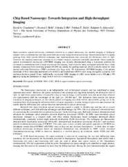
Chip Based Nanoscopy: Towards Integration and High-throughput Imaging
(Journal article; Tidsskriftartikkel; Peer reviewed, 2017)Super-resolution optical microscopy, commonly referred to as optical nanoscopy, has enabled imaging of biological samples with a resolution that was only achievable previously using electron microscopy. Optical nanoscopy is a rapidly growing field, with several different techniques and implementations that overcome the diffraction limit of light. However, the common nanoscope continues to be a rather ... -
Chip-based optical microscopy for imaging membrane sieve plates of liver scavenger cells
(Journal article; Tidsskriftartikkel; Peer reviewed, 2015-08-26)The evanescent field on top of optical waveguides is used to image membrane network and sieve-plates of liver endothelial cells. In waveguide excitation, the evanescent field is dominant only near the surface (~100-150 nm) providing a default optical sectioning by illuminating fluorophores in close proximity to the surface and thus benefiting higher signal-to-noise ratio. The sieve plates of liver ... -
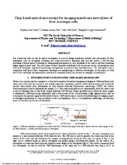
Chip-based wide field-of-view nanoscopy
(Journal article; Tidsskriftartikkel; Peer reviewed, 2017-04-24)Present optical nanoscopy techniques use a complex microscope for imaging and a simple glass slide to hold the sample. Here, we demonstrate the inverse: the use of a complex, but mass-producible optical chip, which hosts the sample and provides a waveguide for the illumination source, and a standard low-cost microscope to acquire super-resolved images via two different approaches. Waveguides composed ... -
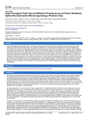
High-Throughput Total Internal Reflection Fluorescence and Direct Stochastic Optical Reconstruction Microscopy Using a Photonic Chip
(Journal article; Tidsskriftartikkel; Peer reviewed, 2019-11-16)Total internal reflection fluorescence (TIRF) is commonly used in single molecule localization based super-resolution microscopy as it gives enhanced contrast due to optical sectioning. The conventional approach is to use high numerical aperture microscope TIRF objectives for both excitation and collection, severely limiting the field of view and throughput. We present a novel approach to generating ... -
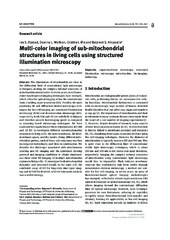
Multi-color imaging of sub-mitochondrial structures in living cells using structured illumination microscopy
(Journal article; Tidsskriftartikkel; Peer reviewed, 2018-05-18)The dimensions of mitochondria are close to the diffraction limit of conventional light microscopy techniques, making the complex internal structures of mitochondria unresolvable. In recent years, new fluorescence-based optical imaging techniques have emerged, which allow for optical imaging below the conventional limit, enabling super-resolution (SR). Possibly the most promising SR and diffraction-limited ... -
New ways of looking at very small holes – using optical nanoscopy to visualize liver sinusoidal endothelial cell fenestrations
(Journal article; Tidsskriftartikkel; Peer reviewed, 2018-01-10)Super-resolution fluorescence microscopy, also known as nanoscopy, has provided us with a glimpse of future impacts on cell biology. Far-field optical nanoscopy allows, for the first time, the study of sub-cellular nanoscale biological structures in living cells, which in the past was limited to electron microscopy (EM) (in fixed/dehydrated) cells or tissues. Nanoscopy has particular utility in the ... -
Photonic-chip assisted correlative light and electron microscopy
(Journal article; Tidsskriftartikkel; Peer reviewed, 2020-12-07)Correlative light and electron microscopy (CLEM) unifies the versatility of light microscopy (LM) with the high resolution of electron microscopy (EM), allowing one to zoom into the complex organization of cells. Here, we introduce photonic chip assisted CLEM, enabling multi-modal total internal reflection fluorescence (TIRF) microscopy over large field of view and high precision localization of the ... -
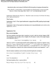
Quantitative phase microscopy of red blood cells during planar trapping and propulsion
(Journal article; Tidsskriftartikkel; Peer reviewed, 2018-08-22)Red blood cells (RBCs) have the ability to undergo morphological deformations during microcirculation, such as changes in surface area, volume and sphericity. Optical waveguide trapping is suitable for trapping, propelling and deforming large cell populations along the length of the waveguide. Bright field microscopy employed with waveguide trapping does not provide quantitative information about ... -
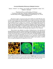
Structured Illumination Microscopy of Biological Structures
(Conference object; Konferansebidrag, 2018)<p>Resolution in optical microscopy has long been limited to the Abbe diffraction limit, i.e. about 250 nm laterally for visible wavelengths on a very good microscope. In the last two decades several techniques have been devised to circumvent this limit: an achievement which was recognized with the 2014 Nobel Prize in Chemistry. Structured Illumination Microscopy (SIM) was the first of these techniques ... -
Uptake and Degradation of Bacteriophages by Liver Sinusoidal Endothelial Cells
(Conference object; Konferansebidrag, 2018)<p>Bacteriophages (briefly, “phages”) are viruses which target bacteria, and are non-infectious to eukaryotic cells. It is estimated that more than 30 billion phages cross into the human body from the gut each day1, and eventually need to be cleared from the blood circulation. The liver plays a central role in pathogen clearance, and liver sinusoidal endothelial cells (LSECs), which form the lining ...